Lung Cancer: A Comprehensive Guide to the Leading Cause of Cancer Death
Introduction: The Global Burden and the Dawn of Precision Medicine
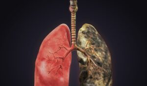
Lung cancer is a relentless global health crisis, responsible for more cancer-related deaths worldwide than breast, prostate, and colorectal cancers combined. Its staggering mortality stems from a deadly combination of high incidence, aggressive biology, and frequent late-stage diagnosis. For decades, the shadow of tobacco use defined this disease, and it remains the paramount preventable cause. However, the past 15 years have witnessed a revolution in lung cancer care, transforming it from a disease with limited, blunt treatment options into a paradigm of precision oncology. The discovery of targetable driver mutations and the advent of immunotherapy have redefined survival expectations and quality of life for thousands of patients. This guide details the complex landscape of lung cancer, from its primary causes and types to the groundbreaking diagnostic and therapeutic strategies that are reshaping its prognosis.
Part 1: Anatomy, Types, and the Critical Distinction
Lung cancers are broadly categorized into two main types, which have profound implications for treatment and prognosis.
1. Non-Small Cell Lung Cancer (NSCLC)
2. Small Cell Lung Cancer (SCLC)
Frequency: ~15% of cases.
Characteristics: Almost exclusively associated with heavy tobacco use. It is a high-grade neuroendocrine tumor characterized by rapid growth, early metastasis, and initial sensitivity to chemotherapy and radiation, but with a high rate of relapse and poor long-term survival. It is rarely treated with surgery.
The “NSCLC vs. SCLC” distinction is the first and most critical fork in the diagnostic and treatment pathway.
Part 2: Etiology and Risk Factors – Beyond Smoking
While tobacco is the dominant cause, the etiology is multifaceted.
Primary Cause:
Other Significant Risk Factors:
Radon Gas: The second leading cause in the U.S. A colorless, odorless radioactive gas from natural uranium decay in soil that can accumulate in homes.
Occupational Exposures: Asbestos (synergistic with smoking), silica, diesel exhaust, arsenic, chromium, and nickel.
Air Pollution: Chronic exposure to particulate matter (PM2.5).
Genetic/Family History: Having a first-degree relative with lung cancer increases risk, suggesting genetic susceptibility.
Previous Radiation Therapy to the chest (e.g., for Hodgkin’s lymphoma).
Scarring from Lung Disease: Such as from tuberculosis or pulmonary fibrosis.
Part 3: Signs and Symptoms – The Challenge of Late Presentation
Early-stage lung cancer is often asymptomatic. When symptoms arise, the disease is frequently advanced.
Symptoms of Primary Tumor:
Persistent or worsening cough (a “new” cough or change in a chronic “smoker’s cough”).
Coughing up blood or rust-colored phlegm (hemoptysis).
Chest pain that worsens with deep breathing, coughing, or laughing.
Hoarseness.
Shortness of breath.
Wheezing.
Symptoms of Locally Advanced Disease:
Symptoms of Metastatic Disease (Distant Spread):
Bone pain (e.g., in back or hips).
Neurological symptoms: Headache, weakness/numbness in limbs, dizziness, seizures (from brain metastases).
Jaundice (yellowing skin/eyes from liver metastases).
Lumps near the surface of the body from metastasis to skin or lymph nodes (e.g., in the neck or above the collarbone).
Paraneoplastic Syndromes: Symptoms caused by hormone-like substances secreted by the tumor, not by the tumor itself (e.g., SIADH, hypercalcemia, clubbing of fingers).
Part 4: Diagnosis and Staging – The Era of Molecular Profiling
Diagnosis requires a tissue biopsy, but modern workup goes far beyond confirming malignancy.
Imaging: A chest X-ray may first detect an abnormality, but a non-contrast chest CT scan is the primary diagnostic tool. PET/CT is then used for staging to identify metabolically active disease in lymph nodes and distant organs.
Biopsy & Pathological Diagnosis: Methods include:
CT-guided needle biopsy (for peripheral lesions).
Bronchoscopy (with EBUS – Endobronchial Ultrasound for central lesions and lymph node sampling).
Surgical biopsy (VATS – Video-Assisted Thoracoscopic Surgery).
The biopsy confirms NSCLC vs. SCLC and the specific subtype.
The Imperative of Molecular (Genomic) Testing (For NSCLC): This is the cornerstone of precision medicine. Testing is done on biopsy tissue (or via liquid biopsy of blood) to identify specific targetable driver mutations and biomarkers:
EGFR, ALK, ROS1, BRAF V600E, KRAS G12C, MET, RET, NTRK: Presence of these mutations/translocations makes the tumor eligible for highly effective oral targeted therapies (TKIs – Tyrosine Kinase Inhibitors).
PD-L1 Expression Level: A protein that helps determine eligibility and likely benefit from immunotherapy.
Next-Generation Sequencing (NGS) panels efficiently test for many alterations simultaneously.
Staging (TNM System for NSCLC):
Stage I & II: Localized to the lung. Potentially curable with surgery.
Stage III: “Locally Advanced.” Tumor involves lymph nodes in the center of the chest (mediastinum). Complex treatment with chemoradiation +/- immunotherapy.
Stage IV: Metastatic. Cancer has spread to the other lung or distant organs. Treated with systemic therapy.
Part 5: Treatment – A Personalized, Evolving Arsenal
Treatment is dictated by cancer type (NSCLC vs. SCLC), stage, molecular profile, and patient health.
A. Treatment for Non-Small Cell Lung Cancer (NSCLC):
B. Treatment for Small Cell Lung Cancer (SCLC):
Limited Stage (confined to one lung and local lymph nodes): Chemotherapy (etoposide + cisplatin/carboplatin) concurrently with thoracic radiation, followed by prophylactic cranial irradiation (PCI) to prevent brain metastases.
Extensive Stage (spread beyond one lung): Immunotherapy (atezolizumab or durvalumab) combined with chemotherapy is the first-line standard. Despite initial response, relapse is common, and second-line options are limited.
Supportive & Palliative Care:
Integrated early, palliative care focuses on managing symptoms (pain, shortness of breath, fatigue), addressing psychological distress, and improving quality of life—and has been shown to extend survival.
Part 6: Screening, Prevention, and the Future
Screening: Annual low-dose CT (LDCT) scan is recommended for high-risk individuals: ages 50-80, with a 20 pack-year smoking history, who currently smoke or have quit within the past 15 years. Screening reduces lung cancer mortality by 20% by detecting cancers at earlier, more curable stages.
Prevention: Tobacco cessation is paramount. Avoidance of radon (home testing) and occupational carcinogens is also critical.
The Future: Research focuses on better screening biomarkers (blood tests), next-generation targeted therapies to overcome resistance, novel immunotherapies, and vaccines for both prevention and treatment.
Conclusion: From Stigma to Hope in the Molecular Era
Lung cancer’s narrative is shifting from one of nihilism to one of cautious optimism. The stigma of a “self-inflicted” disease is being replaced by a scientific understanding of its complex biology. The paradigm has moved from a one-size-fits-all approach to a highly personalized strategy where treatment is selected based on the unique genetic blueprint of an individual’s tumor. While tobacco-related cancers remain a massive challenge, the rise of precision oncology offers unprecedented hope. For patients, this means seeking screening if eligible, demanding comprehensive molecular testing at diagnosis, and accessing care at centers with expertise in thoracic oncology. The battle against lung cancer is being fought with increasingly sophisticated tools, turning a formidable foe into a manageable condition for a growing number of people.
Key Takeaways:
Lung cancer is the leading cause of cancer death, but prevention (smoking cessation) and screening (LDCT) save lives.
Not all lung cancers are the same. Distinguishing NSCLC from SCLC and identifying specific mutations in NSCLC is critical.
Stage IV is no longer an automatic death sentence. Targeted therapies and immunotherapies can control the disease for years.
Molecular testing (genomic profiling) is non-negotiable for every patient with advanced NSCLC.
Palliative care integrated early improves both quality and length of life.
Resources:
Disclaimer: This article is for informational purposes only and is not a substitute for professional medical advice. All treatment decisions must be made in consultation with a multidisciplinary thoracic oncology team.