Bladder Cancer: A Comprehensive Guide to the Industrial and Environmental Cancer
Introduction: The Sentinel of Systemic Exposure
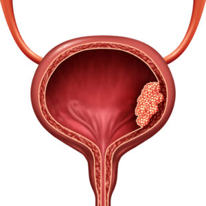
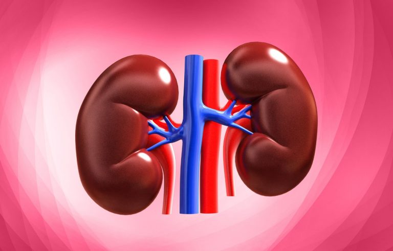
Bladder cancer stands as a unique and telling malignancy, often acting as a biological sentinel for cumulative exposure to environmental and industrial toxins. It is the sixth most common cancer in the United States, with a striking 4:1 male-to-female predominance. The bladder, a hollow muscular organ that stores urine, is lined by a specialized epithelium called the urothelium (or transitional epithelium), which is in constant contact with waste products and potential carcinogens filtered by the kidneys. This direct, prolonged exposure is central to the disease’s etiology. Bladder cancer is characterized by a high rate of recurrence, necessitating vigilant, lifelong surveillance for many patients. However, when confined to the superficial layers, it is highly treatable, while muscle-invasive disease presents a more formidable challenge. This guide details the spectrum of bladder cancer, from non-invasive papillary tumors to aggressive carcinomas, its strong link to smoking and occupational hazards, and the advanced surgical and immunological treatments that define modern care.
Part 1: Anatomy, Types, and the Spectrum of Disease
The bladder wall consists of several layers: the inner urothelium, a supporting lamina propria, the muscularis propria (detrusor muscle), and an outer serosal layer. Cancer staging and treatment hinge on the depth of invasion.
Histological Types:
Urothelial Carcinoma (Transitional Cell Carcinoma – TCC): Accounts for over 90% of bladder cancers in the Western world. Arises from the urothelial cells.
Squamous Cell Carcinoma (~5%): Often associated with chronic irritation, such as from long-term catheter use, bladder stones, or parasitic infection (schistosomiasis).
Adenocarcinoma (~2%): Rare, may arise from urachal remnants or metaplasia.
Small Cell Carcinoma: Very rare, highly aggressive, neuroendocrine origin.
The Clinical Spectrum: A Tale of Two Diseases
Non-Muscle-Invasive Bladder Cancer (NMIBC) – ~75% of new diagnoses:
Confined to the urothelium (Ta: papillary non-invasive) or lamina propria (T1: invasive into connective tissue). Includes Carcinoma In Situ (CIS/Tis), a flat, high-grade lesion that is aggressive despite being non-invasive.
Character: High recurrence rate (50-70%) but low immediate risk of progression to muscle invasion. Requires repeated endoscopic treatments and surveillance.
Muscle-Invasive Bladder Cancer (MIBC) – ~25% of new diagnoses:
Invades into or through the detrusor muscle (T2-T4).
Character: High risk of metastasis and cancer-specific death. Requires aggressive, potentially curative multimodal therapy.
Part 2: Etiology and Risk Factors – The Clear Link to Carcinogens
Bladder carcinogens are absorbed systemically, filtered by the kidneys, concentrated in urine, and stored for hours in the bladder, bathing the urothelium.
Established Risk Factors:
Tobacco Smoking: The #1 risk factor, responsible for ~50% of all cases. Smokers have a 3-4x higher risk. Carcinogens like arylamines (2-naphthylamine) are metabolized and excreted in urine.
Occupational Exposure: Accounts for 15-25% of cases. High-risk industries include:
Dye, rubber, leather, and paint manufacturing.
Truck driving (diesel exhaust).
Hairdressing (aromatic amines in older dyes).
Metalworkers, machinists.
Chemicals: Arylamines, polycyclic aromatic hydrocarbons (PAHs).
Chronic Inflammation: From long-term indwelling catheters, bladder stones, or recurrent urinary tract infections (UTIs), leading to squamous cell carcinoma.
Schistosoma haematobium Infection: A parasitic infection endemic in parts of Africa and the Middle East, strongly linked to squamous cell carcinoma.
Prior Radiation or Chemotherapy: Especially cyclophosphamide.
Arsenic in drinking water.
Age, Gender, and Race: Risk increases with age (median diagnosis 73). Men are far more likely to be diagnosed. White individuals have twice the risk of Black individuals.
Part 3: Signs and Symptoms – Hematuria as the Cardinal Sign
Gross Hematuria (visible blood in urine) is the presenting symptom in 80-90% of patients. It is often painless, intermittent, and may resolve spontaneously, leading to dangerous delays in diagnosis.
Other Symptoms:
Irritative Voiding Symptoms: Urgency, frequency, dysuria (painful urination)—especially common with CIS.
Symptoms of Advanced Disease: Flank pain (from ureteral obstruction), pelvic pain, lower extremity edema, bone pain, weight loss.
Crucial Clinical Rule: Any episode of gross hematuria in an adult over 40 requires a urological evaluation to rule out bladder cancer. Microscopic hematuria also warrants investigation based on risk factors.
Part 4: Diagnosis and Staging – The Urologist’s Toolkit
Diagnosis requires visualizing the inside of the bladder and obtaining tissue.
Cystoscopy: The cornerstone procedure. A thin, flexible scope is passed through the urethra into the bladder.
Blue Light Cystoscopy (with Hexaminolevulinate – Cysview): A photosensitizing agent is instilled, causing cancer cells to fluoresce under blue light, improving detection rates of small or flat tumors (like CIS).
Transurethral Resection of Bladder Tumor (TURBT): The diagnostic and initial therapeutic procedure. Under anesthesia, a resectoscope is used to remove all visible tumor for pathological analysis. This determines:
Histology and Grade (low-grade vs. high-grade).
Stage (Depth of Invasion – Ta, T1, T2+).
Presence of CIS.
Urine Studies:
Cytology: Examines urine for malignant cells. Highly specific for high-grade tumors but insensitive for low-grade.
Molecular Tests (e.g., UroVysion, Cxbladder): Detect genetic or protein markers shed by cancer cells. Used as an adjunct.
Imaging for Staging (for High-Grade/MIBC):
CT Urogram (or MR Urogram): Evaluates the upper urinary tracts (kidneys, ureters) for tumors and assesses for local invasion and lymph node involvement.
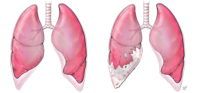
CT Chest/Abdomen/Pelvis: For metastatic workup.
Bone Scan: If bone pain or elevated alkaline phosphatase is present.
Part 5: Treatment – A Risk-Stratified, Stepwise Approach
A. Treatment for Non-Muscle-Invasive Bladder Cancer (NMIBC):
Management is dictated by risk stratification (based on grade, stage, size, multifocality, and presence of CIS).
Low-Risk NMIBC: TURBT alone, followed by surveillance cystoscopy.
Intermediate & High-Risk NMIBC:
Complete TURBT.
Intravesical Therapy: Medication instilled directly into the bladder via a catheter.
Chemotherapy (e.g., Mitomycin C, Gemcitabine): Used for immediate post-TURBT instillation or for intermediate-risk disease to reduce recurrence.
Immunotherapy – BCG (Bacillus Calmette-Guérin): The gold standard for high-risk NMIBC and CIS. A live, attenuated bacterium stimulates a potent local immune response against cancer cells. Given as a 6-week induction course followed by maintenance therapy.
For BCG-Unresponsive NMIBC: Options include novel intravesical agents (e.g., nadofaragene firadenovec gene therapy, immunotherapy), or radical cystectomy.
B. Treatment for Muscle-Invasive Bladder Cancer (MIBC):
The goal is cure. Requires a multidisciplinary team.
Radical Cystectomy with Pelvic Lymph Node Dissection: The historical gold standard. Removal of the entire bladder, surrounding tissues, and pelvic lymph nodes.
Trimodal Therapy (TMT) / Bladder Preservation: An alternative for select patients.
Neoadjuvant Chemotherapy: Administration of cisplatin-based chemotherapy BEFORE cystectomy is the standard of care for eligible patients. It improves overall survival by treating micrometastatic disease.
Adjuvant Chemotherapy: Given after cystectomy if no neoadjuvant therapy was given and pathology shows high-risk features.
C. Treatment for Advanced/Metastatic Disease:
First-Line Therapy: Platinum-based chemotherapy (e.g., gemcitabine/cisplatin) remains foundational.
Maintenance Therapy: For patients who respond to first-line chemo, switching to avelumab (an immunotherapy) maintenance improves survival.
Second-Line & Beyond – The Immunotherapy Revolution:
Targeted Therapy: Erdafitinib for tumors with specific FGFR genetic alterations.
Antibody-Drug Conjugates: Enfortumab vedotin (targets Nectin-4) and Sacituzumab govitecan (targets Trop-2) have shown impressive activity in later lines.
Part 6: Prognosis and the Imperative of Surveillance
Prognosis: For NMIBC, 5-year survival is >90%, but recurrence is the rule, not the exception. For MIBC, 5-year survival is ~50-60% with optimal treatment, but drops sharply with nodal or distant metastasis.
Surveillance: Is lifelong and intensive, especially for NMIBC, involving regular cystoscopies and imaging of the upper tracts due to risk of recurrence in the bladder and ureters/renal pelvis (field carcinogenesis).
Survivorship Issues: Significant impact on quality of life from urinary diversion, sexual dysfunction post-cystectomy, and the psychological burden of constant surveillance.
Conclusion: A Model of Chronic Cancer Management and Innovation
Bladder cancer epitomizes the concept of a “chronic cancer,” where long-term management of recurrence is balanced against the threat of progression. Its strong environmental links make it a preventable disease through smoking cessation and workplace safety. The treatment landscape is one of remarkable innovation, from the century-old immune stimulant BCG to cutting-edge antibody-drug conjugates and targeted therapies for metastatic disease. For patients, success hinges on prompt evaluation of hematuria, adherence to rigorous surveillance protocols, and access to a specialized urologic oncology team capable of navigating the complex decisions from intravesical therapy to radical cystectomy and systemic treatment. Through prevention, early detection, and personalized therapy, the significant morbidity and mortality of bladder cancer can be steadily reduced.
Key Takeaways:
Painless blood in the urine is bladder cancer until proven otherwise. See a urologist immediately.
Smoking is the leading preventable cause. Quitting reduces risk.
NMIBC has a high recurrence rate but is rarely life-threatening if managed properly with TURBT and intravesical therapy (like BCG).
MIBC is a serious disease requiring aggressive, potentially curative treatment (cystectomy or chemoradiation).
Surveillance is a lifelong commitment for most patients.
Resources:
Disclaimer: This article is for informational purposes only and is not a substitute for professional medical advice, diagnosis, or treatment. Always seek the advice of a urologic oncologist with any questions you may have regarding a medical condition.