Squamous Cell Carcinoma: A Comprehensive Guide to the Second Most Common Skin Cancer
Introduction: The Sun’s Cumulative Debt
Squamous cell carcinoma (SCC) is a pervasive and potentially serious form of skin cancer that arises from the uncontrolled growth of squamous cells in the epidermis—the skin’s outermost layer. As the second most common skin cancer, with over 1.8 million cases diagnosed annually in the U.S., it represents a significant public health burden. Unlike its cousin, basal cell carcinoma, which is rarely fatal, SCC carries a measurable risk of metastasis (spread) and can be life-threatening if neglected. SCC is the definitive consequence of cumulative, lifelong sun exposure, making it a disease of chronic photodamage. While highly curable when detected early, it underscores the critical importance of sun protection and vigilant skin surveillance. This guide provides a detailed exploration of SCC’s causes, identification, advanced risk factors, and modern treatment strategies.
Part 1: The Biology and Pathogenesis of SCC
Squamous cells are flat, scale-like cells that make up most of the epidermis. SCC develops when DNA damage in these cells—primarily from ultraviolet (UV) radiation—triggers mutations that lead to uncontrolled replication.
The Carcinogenic Pathway: UVB rays directly damage cellular DNA, creating “signature mutations.” UVA rays generate reactive oxygen species, causing indirect damage. When the body’s repair mechanisms fail (a process more likely with age and immunosuppression), mutated cells begin to proliferate.
Precancerous Lesions: SCC often develops from premalignant precursors, serving as critical warning signs:
Actinic Keratosis (AK): The most common precursor. Rough, scaly, sandpaper-like patches on sun-exposed skin. Only a small percentage progress to SCC, but their presence indicates significant sun damage and increased cancer risk.
Bowen’s Disease (Squamous Cell Carcinoma in situ): An early, non-invasive form of SCC confined to the epidermis. Appears as a persistent, slowly enlarging, red, scaly patch.
Progression: From AK, it can progress to invasive SCC, which penetrates through the basement membrane into the dermis. Once invasive, it gains the potential to metastasize, typically first to nearby lymph nodes.
Part 2: Risk Factors: Beyond Simple Sun Exposure
While UV exposure is the paramount cause, other factors significantly modulate risk.
Primary Risk Factors:
Cumulative UV Exposure: Lifetime sun exposure is the strongest predictor. Occupational and recreational sun exposure, sunburns (especially in childhood), and tanning bed use (“indoor tanning” is a Class I carcinogen) are major contributors.
Skin Phenotype: Fair skin (Fitzpatrick Types I-II), light eyes, red or blond hair, and a tendency to freckle or burn easily.
Age: Risk increases dramatically after age 50, reflecting the time required for cumulative DNA damage.
Gender: Men are 2-3 times more likely to develop SCC, often due to greater occupational sun exposure and lower rates of sun protection use.
Geography: Living closer to the equator or at high altitudes with more intense UV radiation.
Major Risk Amplifiers:
Immunosuppression: This is a critical risk multiplier. Organ transplant recipients (on lifelong immunosuppressive drugs) have a 65-250 times higher risk of developing SCC, which also tends to be more aggressive and numerous. Other conditions include HIV/AIDS and hematologic malignancies.
Chronic Inflammation or Injury: SCC can arise in long-standing scars (from burns, trauma), chronic ulcers, sinus tracts, and sites of previous radiation therapy.
Human Papillomavirus (HPV) Infection: Certain beta-genus HPV types (especially in immunocompromised individuals) are linked to SCC, particularly in the anogenital region and around fingernails.
Chemical Carcinogen Exposure: Long-term exposure to arsenic (historically in well water or pesticides) and industrial hydrocarbons.
Part 3: Clinical Presentation – Recognizing the Lesion
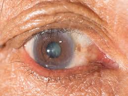
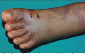
Early recognition is key. SCC can appear on any sun-exposed area but is most common on the rim of the ear, face (especially lips, nose, and scalp in balding men), neck, hands, and arms.
Common Appearances:
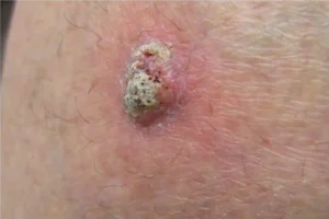
Persistent, Scaly Red Patch: With irregular borders, may crust or bleed.
Elevated Growth with a Central Depression: Often resembles a wart or a volcano-like nodule. The surface may be crusted or ulcerated.
Open Sore (Ulcer): That does not heal, or heals and re-opens, over weeks to months.
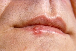
A Firm, Red Nodule: On the lip or ear, which can be particularly high-risk locations.
An Old Scar or Ulcer: That develops new growth, tenderness, or ulceration.
“High-Risk” Features That Demand Urgent Attention:
Diameter > 2 cm (≈ size of a nickel).
Location on the lip, ear, scalp, genitals, or hands.
Rapid growth.
Recurrence of a previously treated lesion.
Symptoms like pain, tenderness, or bleeding.
Depth of invasion > 2 mm or invasion into subcutaneous fat.
Poor differentiation on biopsy (cells look very abnormal).
Perineural invasion (cancer cells tracking along nerves).
Part 4: Diagnosis and Staging
Diagnosis begins with a skin biopsy, which is mandatory for any suspicious lesion.
Shave, Punch, or Excisional Biopsy: The dermatologist removes all or part of the lesion for pathological examination. The biopsy report will confirm SCC and detail critical prognostic factors: depth (Breslow thickness), grade (differentiation), perineural invasion, and margin status.
Staging: For most localized, low-risk SCCs, no further staging is needed after complete excision. For high-risk SCCs, staging workup may include:
Imaging: MRI (if perineural invasion is suspected), CT, or PET/CT to check for spread to lymph nodes or distant organs.
Sentinel Lymph Node Biopsy (SLNB): An emerging consideration for the highest-risk tumors, though not yet standard.
Part 5: Treatment Modalities – From Cure to Control
Treatment is highly effective for early-stage SCC and is chosen based on tumor size, location, risk features, and patient health.
Primary Curative Treatments (for localized SCC):
Surgical Excision: The gold standard. The tumor is cut out with a margin of normal-appearing skin (typically 4-6 mm for low-risk, wider for high-risk). The specimen is sent to pathology to ensure clear margins—no cancer cells at the edges.
Mohs Micrographic Surgery: The treatment of choice for high-risk SCC and tumors in cosmetically or functionally sensitive areas (face, ears, lips, genitals).
Process: The surgeon removes thin layers of tissue, which are immediately mapped, frozen, and examined under a microscope during the surgery. This continues until a cancer-free plane is reached.
Advantage: Maximizes cure rate (up to 99% for primary tumors) while preserving the maximum amount of healthy tissue.
Other Treatment Options:
Electrodessication and Curettage (ED&C): Suitable only for small, thin, low-risk SCCs on the trunk or extremities. Involves scraping away the tumor and cauterizing the base.
Radiation Therapy: An excellent option for patients who are not surgical candidates, for tumors in locations where surgery is difficult, or as adjuvant therapy after surgery for high-risk features (e.g., perineural invasion).
Topical Therapies: For SCC in situ (Bowen’s disease) or very superficial SCCs. Options include 5-fluorouracil (5-FU) cream, imiquimod cream, or photodynamic therapy (PDT).
Treatment for Advanced, Metastatic, or Inoperable SCC:
This is a rapidly evolving field.
Immunotherapy: PD-1 Inhibitors (Cemiplimab, Pembrolizumab) have revolutionized treatment. They work by blocking a cancer cell’s ability to “hide” from the immune system, allowing the body’s T-cells to attack the tumor. They are now first-line for metastatic or locally advanced SCC.
Targeted Therapy: EGFR Inhibitors (Cetuximab) may be used, often in combination with radiation or chemotherapy.
Chemotherapy: Traditional agents like cisplatin may be used, but are generally less effective than immunotherapy.
Part 6: Prevention and Prognosis
Prevention is Paramount:
Daily Sun Protection: Broad-spectrum SPF 30+ sunscreen on all exposed skin, year-round. Reapply every 2 hours.
Protective Clothing: Wide-brimmed hats, UV-protective clothing, sunglasses.
Seek Shade: Especially between 10 a.m. and 4 p.m.
Avoid Tanning Beds Entirely.
Regular Skin Self-Exams: Know your skin and report any new, changing, or non-healing spots to a dermatologist immediately.
Full-Body Skin Exams: Annual professional exams for high-risk individuals (fair skin, history of skin cancer, immunosuppression).
Prognosis:
The cure rate for early, localized SCC treated appropriately is over 95%.
Metastatic SCC has a poorer prognosis, but the advent of immunotherapy has significantly improved survival rates.
A history of one SCC significantly increases the risk of developing another, emphasizing the need for lifelong vigilance.
Conclusion: A Clear and Present Danger, a Manageable Foe
Squamous cell carcinoma is a direct and tangible consequence of our interaction with the sun. It serves as a biological receipt for a lifetime of UV exposure. While common, it should never be considered trivial due to its metastatic potential. The modern management of SCC is a success story of prevention through sun safety, early detection through surveillance, and precise, effective treatment. For patients, this means adopting a proactive partnership with a dermatologist: practicing rigorous sun protection, performing regular self-exams, and seeking prompt evaluation for any suspicious lesion. Through this combination of personal diligence and advanced medicine, the significant burden of SCC can be dramatically reduced.
When to See a Dermatologist:
Any new, growing, or changing skin growth.
A sore that does not heal within 4-6 weeks.
Any rough, scaly patch (actinic keratosis) that becomes tender, raised, or ulcerated.
A history of skin cancer or significant sun damage.
Disclaimer: This article is for informational purposes only and is not a substitute for professional medical advice, diagnosis, or treatment. Always seek the advice of a board-certified dermatologist with any questions you may have regarding a skin lesion or medical condition.